
Trinocular Polarizing Microscope 80%/20% beam-splitter with a large stable microscope base and limb with graduated coaxial coarse and fine focusing (with safety stop),rack and pinion focusing condenser mount.
Equipped with strain free condenser and 4X, 10X, 40XS strain free objectives with pre-centered transmitted and incident 6V 30W light intensity Halogen lamp with variable intensity control
Features include
- DIN standard eyepiece HWF10X
- DIN focusing HWF10XF eyepiece with cross-line reticle
- Centerable quadruple nosepiece
- SM Plan Strain Free objectives 4X, 10X,40X(S) (Magnification factor 1.5X)
- Rotatable stage with graduation 360° with vernier
- Strain Free Abbe condenser NA 1.25 with Iris, filter holder and blue filter
- Rotatable polarizer in swing-out mount, analyzer and Bertrand lens in sliding mount
- Coaxial coarse and fine adjustment with graduation
- Built-in Koehler illuminator with halogen lamp 6V 30W light intensity control
- Mica 1/4 wave plate
- First order red plate
- Slide opening in top limb for insertion of DIN standard 20mm X 6mm compensator, slide away SE-NW (Mica 1/4 wave plate and sensitive tint plate included)
Brightfield
The simplest of all the optical microscopy illumination techniques. Sample illumination is transmitted (i.e., illuminated from below and observed from above) white light and contrast in the sample is caused by absorbance of some of the transmitted light in dense areas of the sample. Brightfield microscopy is the simplest of a range of techniques used for illumination of samples in light microscopes and its simplicity makes it a popular technique. The typical appearance of a brightfield microscopy image is a dark sample on a bright background.
Polarizing Light
A type of optical microscopy techniques involving polarized light. Simple techniques include illumination of the sample with polarized light. Directly transmitted light can, optionally, be blocked with a polarizer orientated at 90° degrees to the illumination. More complex microscopy techniques which take advantage of polarized light include differential interference contrast microscopy and interference reflection microscopy.
Applications
- Environmental Research - Analysis of materials, dust and fiber
- Medicine and Biology - Determining composition of calculi, sediments and bio-crystallates
- Forensics - Identification of trace elements and materials such as explosives
- Materials Analysis - Metal, ceramics, glass, building materials, etc.
- Geology - Mineralogy, petrography, core drilling sample analysis, thin section identification
- Chemistry - Quality control of in-process or end products, compounds, pharmaceutical analysis and identification of precipitates
- Weight: 31.0 lbs (8.5 kg)
- Box Size: 22 x 16 x 14 inches
- Footprint: 254mm (D) x 203mm (W)
- Trinocular head type: 270 mm(D) x 480 mm (H) x 200mm(W) 32.0 lbs. (8.7 kg)
| Model | Head | Illumination | Eyepieces | Objectives | Condenser | Stage |
ML9430 | Trinocular | Both 6V 30W halogen vertical Koehler and 6V 30W halogen transmitted Koehler | HWF10x and HWF10x w/crossline | Strain-free SM. Plan 4x, 10x, S40x | Strain-free NA 1.25 achromatic w/ iris diaphragm and rack and pinion focusing | Rotatable, centerable, circular stage w/clips |
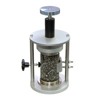
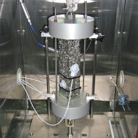
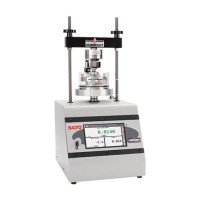
Uniaxial Fatigue (S-VECD) Test for UTM, AsphaltQube, AMPT Pro and AMPTQube
Controls - 79-PV70600, 79-PV70610, 79-PV70611, 79-PV70619
Upon Request
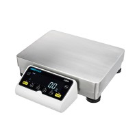
HIGH CAPACITY PRECISION BALANCCES WITH BELLOW MEASUREMENT
Geneq - LBL14001, LBL24001, LBL34001, LBL14001P, LBL24001P, LBL34001P
Upon Request
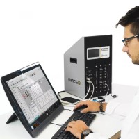
UTS Neutron – Dynamic Materials Testing & Analysis Software
Controls - 79-UTS2000, 79-UTS2100, 79-UTS2200
Upon Request

Can we help you?
min 10 ch